
ishihara 14 plate test pdf
Ishihara 14 Plate Test PDF: A Comprehensive Guide

This guide details the Ishihara 14-plate test, a quick assessment for red-green color vision deficiencies, readily available as a PDF for convenient access and use.
What is the Ishihara Test?
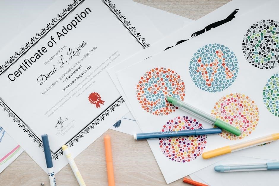
The Ishihara Test is a widely utilized diagnostic tool designed to detect deficiencies in color vision, specifically those related to red and green hues. Developed initially in 1917 by Dr. Shinobu Ishihara, a Japanese ophthalmologist, the test employs a series of plates, each displaying a pattern of colored dots.
Embedded within these dot patterns are numbers or shapes formed by dots of a slightly different color. Individuals with normal color vision can easily discern these figures, while those with color vision deficiencies struggle to identify them, or perceive entirely different numbers.
The test comes in various formats, most notably the 38-plate and 14-plate versions. PDF versions of these tests are commonly found online, offering a digital means of screening. The core principle remains consistent: assessing the ability to differentiate between colors to identify potential color blindness.
History and Development of the Ishihara Test
The Ishihara Test originated from the work of Dr. Shinobu Ishihara, who sought a more practical method for identifying color vision deficiencies, particularly within the Japanese military in 1917. Existing tests at the time were cumbersome and less efficient.
Dr. Ishihara’s innovation lay in creating plates with patterns of colored dots concealing numbers or shapes. This allowed for a rapid assessment of an individual’s ability to distinguish between red and green, the most common forms of color blindness. The initial test comprised a set of 38 plates.
Over time, the test gained international recognition and became a standard in ophthalmological and optometric examinations. PDF versions emerged with the advent of digital technology, facilitating wider accessibility and ease of administration. The 14-plate version offers a shorter screening option, while retaining diagnostic value.
Understanding Color Blindness (Color Vision Deficiency)
Color blindness, more accurately termed color vision deficiency, isn’t typically complete blindness to color; rather, it’s a reduced ability to perceive certain colors, most commonly red and green. This condition arises from a malfunction in the cone cells within the retina, responsible for color detection.
These cone cells come in three types, each sensitive to different wavelengths of light – red, green, and blue. A deficiency in one or more of these cone types leads to difficulty distinguishing between certain hues. The Ishihara 14 plate test PDF is a valuable tool in initially identifying these deficiencies.
It’s crucial to understand that color vision deficiency is often genetic, affecting males more frequently than females. While there’s no cure, understanding the type and severity helps individuals adapt and navigate a world designed for full color vision. Early detection, aided by tests like Ishihara, is key.
Types of Color Blindness Detected by the Ishihara Test
The Ishihara test, including the 14-plate PDF version, primarily screens for red-green color vision deficiencies, the most prevalent forms of color blindness. These deficiencies fall into several categories.
Protanopia is a complete absence of red cone cells, while protanomaly involves a reduced sensitivity to red. Similarly, deuteranopia signifies a lack of green cones, and deuteranomaly indicates reduced green sensitivity. The test helps differentiate between these.
Individuals with protanopia and deuteranopia struggle to distinguish between reds, greens, and yellows, often confusing them. Those with protanomaly and deuteranomaly have milder difficulties. The patterns within the Ishihara plates are designed to be easily visible to those with normal color vision but challenging for those with these deficiencies, revealing the specific type based on error patterns.
Prevalence of Color Blindness
Color vision deficiency, detectable using tools like the Ishihara 14-plate test PDF, affects a significant portion of the male population. Approximately 8% of males experience some form of color blindness, a considerably higher rate than females, where the prevalence is around 0.5%.
This disparity is linked to the genetic basis of the most common red-green color blindness, which is X-linked recessive. Males, having only one X chromosome, are more susceptible to inheriting the affected gene. While less common, color blindness can also occur in females, typically requiring inheriting the gene on both X chromosomes.
Globally, millions are affected, highlighting the importance of accessible screening tools like the Ishihara test. Early detection, facilitated by PDF versions, is crucial for individuals pursuing careers where accurate color perception is essential.

The 14-Plate Ishihara Test: Specifics
The 14-plate version offers a concise screening for red-green deficiencies, utilizing dot patterns to reveal numbers; a PDF format enhances accessibility and ease of use.
Purpose of Using a 14-Plate Version
The 14-plate Ishihara test serves as a streamlined, efficient screening tool for color vision deficiencies, particularly red-green color blindness. While the full 38-plate test provides a more comprehensive evaluation, the 14-plate version is often preferred for initial assessments due to its brevity and practicality.
This shorter format is ideal for situations where a quick indication of potential color vision issues is needed, such as preliminary school screenings, pre-employment checks in certain professions, or initial evaluations in clinical settings. A PDF version of the 14-plate test further enhances its usability, allowing for easy distribution, printing, and administration.
It’s a cost-effective and time-saving method to identify individuals who may require further, more detailed testing with the complete 38-plate set or other specialized color vision assessments. The 14-plate test doesn’t definitively diagnose the type of color blindness, but flags potential concerns for further investigation.
How the 14-Plate Test Differs from the 38-Plate Test
The primary difference between the Ishihara 14-plate and 38-plate tests lies in their scope and diagnostic detail. The 38-plate version offers a significantly more thorough assessment of color vision, capable of identifying a wider range of color vision deficiencies and differentiating between various types with greater accuracy.
The 14-plate test, while effective as a screening tool, focuses primarily on detecting the most common forms of red-green color blindness. It provides a quicker, less exhaustive evaluation. A PDF format doesn’t alter this fundamental difference; it simply provides a digital means of administering either test.
Consequently, the 38-plate test is preferred when a precise diagnosis is required, or when assessing individuals for professions where subtle color distinctions are critical. The 14-plate test is suitable for initial screening, but positive results should be followed up with the more comprehensive 38-plate assessment.
Administering the 14-Plate Ishihara Test
Proper administration of the Ishihara 14-plate test, whether using a physical set or a PDF version, is crucial for accurate results. Begin by ensuring the examinee understands the task: to identify the numbers or shapes concealed within the colored dots on each plate;
The test should be conducted individually. Avoid any prompting or assistance during the examination. Present each plate briefly – typically for just a few seconds – to allow for spontaneous responses. Record each answer carefully, noting any hesitation or difficulty the examinee experiences.
Consistency in testing conditions is vital. Maintaining standardized lighting and viewing distance ensures reliable outcomes. A PDF version offers portability, but the screen’s calibration should be checked to ensure accurate color representation.
Proper Lighting Conditions
Optimal lighting is paramount when administering the Ishihara 14-plate test, regardless of whether utilizing physical plates or a PDF version displayed on a screen. The ideal illumination mimics daylight – a neutral, even light source is essential. Avoid strong, direct sunlight or artificial lights with a noticeable color cast, as these can distort color perception and lead to inaccurate results;
The testing area should be well-lit, but not glaring. A matte finish on surfaces minimizes reflections that could interfere with viewing the plates. When using a PDF, ensure the screen isn’t overly bright or dim, and calibrate it for accurate color reproduction.
Consistent lighting throughout the test is critical. Fluctuations can affect the examinee’s ability to discern the hidden figures. Maintaining these conditions ensures a reliable and valid assessment of color vision.
Distance and Viewing Angle
Maintaining a standardized distance and viewing angle is crucial for accurate results when administering the Ishihara 14-plate test, whether using physical plates or a PDF displayed digitally. Typically, the plates should be viewed from a distance of approximately 38-40 centimeters (around 15-16 inches). This distance ensures optimal visual acuity and minimizes distortion.
The examinee should position themselves directly in front of each plate, avoiding any oblique viewing angles. A slight head tilt is permissible for comfort, but significant angles can alter color perception. When using a PDF, ensure the screen is positioned at the correct distance and that the examinee is seated comfortably.
Consistent distance and angle across all plates are vital for reliable comparison of responses. Any deviation can introduce error and compromise the validity of the color vision assessment.

Interpreting the Results of the 14-Plate Test
Analyzing errors on the Ishihara 14-plate test, often accessed as a PDF, reveals the type of color vision deficiency, if present, through a defined scoring system.
Scoring System and Interpretation
The Ishihara test, frequently utilized in its PDF format, employs a straightforward scoring method. Each plate is designed to reveal a specific number to individuals with normal color vision. The test administrator records the number correctly identified by the examinee on each plate. A complete absence of errors indicates normal color vision.
However, the interpretation isn’t simply about the total number of errors. The pattern of errors is crucial. Consistent misidentification of numbers, or an inability to discern any number at all on certain plates, points towards specific types of color vision deficiencies. For example, difficulty seeing numbers on plates designed to test for protanopia or deuteranopia (red-blindness or green-blindness) suggests a deficiency in those color perceptions.
A PDF version of the test often includes guidelines for interpreting the results, but professional assessment is always recommended for a definitive diagnosis. The scoring helps categorize the severity of the deficiency, ranging from mild to severe, aiding in understanding the impact on daily life.
Identifying Different Types of Color Blindness Based on Errors
Analyzing error patterns on the Ishihara 14-plate test, often accessed as a convenient PDF, is key to pinpointing the specific type of color vision deficiency. Consistent mistakes on certain plates suggest particular deficiencies. For instance, difficulty with plates showing red and green hues often indicates deuteranomaly (reduced green sensitivity) or protanomaly (reduced red sensitivity).
Protanopia (red-blindness) and deuteranopia (green-blindness) typically result in complete inability to identify numbers on specific plates. Tritanopia (blue-blindness), though rarer, presents a different error profile. The PDF test’s plates are designed to differentiate these conditions.
It’s important to note that the 14-plate version offers a preliminary assessment. A thorough diagnosis requires professional evaluation. The pattern of errors, combined with a patient’s history, allows clinicians to accurately categorize the type and severity of color blindness, guiding appropriate advice and support.
Common Errors and What They Indicate
When using the Ishihara 14-plate test, often found as a downloadable PDF, certain errors frequently occur and offer clues about potential color vision deficiencies. Misidentifying ‘2’ as ‘3’ or vice versa on specific plates is a common indicator of deuteranomaly, a reduced sensitivity to green light.
Confusing numbers altogether, or seeing no number at all, suggests a more severe deficiency like deuteranopia or protanopia (red-blindness). Difficulty distinguishing shades of red and green is a hallmark of these conditions. The PDF format allows for repeated testing to confirm these observations.
However, it’s crucial to remember that errors can also stem from poor lighting or viewing conditions. A professional assessment is vital for accurate diagnosis. The Ishihara test PDF is a screening tool, not a definitive diagnostic instrument, and consistent errors warrant further investigation.

Ishihara Test PDF: Accessing and Using Digital Resources
Numerous websites offer Ishihara 14-plate test PDFs for download, providing convenient access to this color vision assessment tool for preliminary screening purposes.
Finding Reliable Ishihara 14 Plate Test PDFs Online
Locating a trustworthy Ishihara 14-plate test PDF requires careful consideration, as the internet hosts varying levels of quality and accuracy. Prioritize sources affiliated with reputable organizations, such as optometry practices, vision research institutions, or established medical publishers. Be wary of websites offering free downloads without clear authorship or validation, as these may contain inaccuracies or altered images impacting test results.
Look for PDFs that explicitly state the edition and plate number (14-plate in this case) to ensure you’re using the correct version. Cross-reference the PDF with known examples of genuine Ishihara plates to verify color accuracy and image clarity. Some websites may require registration or a small fee to access high-quality PDFs, which can be a reasonable trade-off for reliability. Always download from secure (HTTPS) websites to protect your device from potential malware. Remember, an online test is a screening tool and should not replace a professional eye examination.
Benefits of Using a PDF Version
Employing an Ishihara 14-plate test PDF offers several practical advantages over other testing methods. PDFs provide immediate accessibility, allowing for quick screening without requiring specialized equipment or a visit to a clinic. They are easily shareable, facilitating preliminary assessments in remote settings or for individuals unable to access professional care easily.
The digital format enables convenient storage and retrieval, eliminating the risk of lost or damaged physical test charts. PDFs can be viewed on various devices – computers, tablets, and smartphones – enhancing portability and usability. Furthermore, a PDF version often allows for printing, creating a tangible copy for repeated use or record-keeping. However, it’s crucial to ensure the PDF is high-resolution to maintain color accuracy, and remember that a PDF test serves as a screening tool, not a definitive diagnosis, necessitating professional confirmation.
Limitations of Online Tests vs. Professional Administration
While Ishihara 14-plate test PDFs and online versions offer convenience, they possess inherent limitations compared to professional administration. Color accuracy is paramount, and screen calibration varies significantly across devices, potentially skewing results. Online tests lack the controlled lighting conditions and standardized viewing distances crucial for reliable assessment.
A trained professional can observe the examinee’s responses and identify subtle errors or hesitation, providing valuable qualitative data. They can also account for individual variations in perception and rule out other potential causes of color vision difficulties. Self-administered online tests are susceptible to user error, intentional or unintentional, impacting accuracy. Therefore, a positive result from an online or PDF test should always be confirmed by a qualified eye care professional for a definitive diagnosis and appropriate guidance.
Beyond the 14-Plate Test: Further Evaluation
If the Ishihara 14-plate test PDF results are inconclusive, or indicate a deficiency, additional, more comprehensive color vision assessments are often necessary for accurate diagnosis.
When Additional Testing is Necessary
While the Ishihara 14-plate test PDF serves as a valuable initial screening tool, it isn’t always definitive. Further evaluation becomes crucial when results are ambiguous, or when a suspected color vision deficiency significantly impacts an individual’s daily life or profession.
Specifically, if an individual struggles to identify patterns on several plates, or consistently misinterprets numbers, more in-depth testing is warranted. Certain professions – pilots, electricians, and designers, for example – require stringent color vision standards, necessitating a more precise diagnosis than the 14-plate test can provide.
Furthermore, the Ishihara test primarily detects red-green color deficiencies; it may not identify all types of color blindness. Therefore, individuals with suspected blue-yellow color blindness, or those with atypical responses on the Ishihara test, should undergo additional assessments to ensure a complete and accurate evaluation of their color vision capabilities. A comprehensive examination by an eye care professional is always recommended.
Other Color Vision Tests Available
Beyond the Ishihara 14-plate test PDF, a range of more sophisticated color vision assessments exist, offering a more detailed and nuanced understanding of an individual’s color perception. These tests are often employed when the Ishihara test yields inconclusive results or when a comprehensive evaluation is required.
The Farnsworth Munsell 100 Hue Test requires arranging colored caps in order of subtle hue changes, revealing deficiencies beyond red-green discrimination. Anomalous Trichromacy can be identified with this test. Another option is the Farnsworth-Hayden Test, designed specifically for detecting blue-yellow deficiencies.
Additionally, color matching functions assess an individual’s ability to match colors, providing quantitative data on their color perception. Cone contrast tests directly measure the function of the cone photoreceptors in the retina. These tests, administered by qualified eye care professionals, provide a more precise diagnosis and guide appropriate management strategies for individuals with color vision deficiencies.